Fitness
‘Extreme’ Cells Could Provide New Insights into Cell Biology, Pregnancy Diseases, and Cancer

The textbook model of a cell with a single nucleus accurately describes most cells — but not all. In fact, many cells have more than one nucleus. The outermost layer of the human placenta is one giant cell with billions of nuclei. Laid flat, it would measure 12-14 square meters.
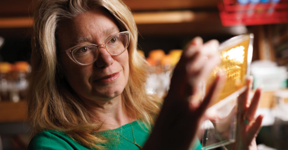
Amy Gladfelter, PhD’01, has been fascinated by such cells for decades. “As soon as I looked under a microscope at these very large fungal cells, I couldn’t stop thinking about them,” she said. “There were so many surprises and paradoxes because most of the rules of cell biology had been established in simple systems, and these cells just kept breaking the rules.”
A professor in the School of Medicine’s Department of Cell Biology, Gladfelter joined the Duke faculty last year as a Duke Science and Technology Scholar.
In her lab, she studies both marine fungi and placental cells. “We’re interested in how being able to take on multinucleated organization and become quite large may help cells cope with dynamic and extreme environments,” she said.
Gladfelter’s work could refine models of cell biology, explain some diseases of pregnancy, and even open up new avenues for treating cancer.
Exploring the Secrets of the Human Placenta
The giant cell that makes up the outermost layer of the placenta has to juggle many conflicting signals related to the needs of the growing fetus and the mother. Gladfelter is exploring how the cell’s size and organization may help it interpret and act on different signals to guide its behavior in the moment.
She’s excited about the work because the placenta’s giant cell has not yet been well described in terms of its organization and function. “We’re going to be understanding how the cell in the placenta works in a healthy pregnancy and what that might teach us about pregnancy diseases where there is not a lot known about what is going on at the cellular level,” she said.
She studies cells with microscopes, analyzing the images with machine-learning techniques and modeling the data to better understand the cells’ behavior. “For us, images are far more than a pretty picture,” she said. “They are a place we extract a lot of information about the dynamics and physical structure of the cells.”
A deeper understanding of the placenta could shed light on cancer, given the intriguing similarities between the two: both seek to evade the immune system, both make use of an unusual type of metabolism, and both upregulate some of the same genes. And cancer cells often have multiple nuclei.
Basic Science + Collaboration = Medical Applications
Gladfelter’s investigations of marine fungi and the giant placenta cell are fueled by her curiosity to understand on a molecular level how these unusual cells function: how they move, how they respond to signals and stress, how they sense their own shapes. And she wants to use that information to improve and expand the current understanding of the inner workings of all cells. “Because these cells are so extreme, they often reveal features that all cells are using,” she said. “They are magnified because of the massive size of the cells.”
Gladfelter has no doubt that basic science is the first step to developing new medical therapies and other technological advances.
“We don’t really begin to have enough of an understanding to solve all the problems that exist right now,” she said. “There are just major gaps in our basic science knowledge that are holding back medical applications.”
She is collaborating with colleagues in the Division of Maternal-Fetal Medicine, the Department of Biomedical Engineering, and the Tisch Brain Tumor Center, among others, to share what she’s discovering with others who are seeking to translate new discoveries into new therapies.
The opportunity for collaboration was part of the reason she jumped at the chance to return to Duke as a faculty member, after having earned her PhD here in 2001. “The openness of people to collaboration was a big piece of it,” she said. “I felt like I could really make interdisciplinary connections here.”
Story originally published in DukeMed Alumni News, Spring 2024.
Read more from DukeMed Alumni News


